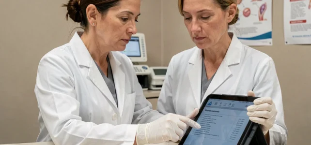
When a cervical smear reveals abnormal cells classified as HSIL (high-grade squamous intraepithelial lesion), the next steps may seem unclear. This result means that cells in the cervix show marked changes related to a persistent HPV infection. These abnormalities are not cancer, but they can lead to it if left untreated. Understanding treatment options allows for a clearer approach to management.
Cervical Conization: The Standard Treatment for HSIL
Conization remains the most commonly used technique for confirmed high-grade lesions. The principle is simple: remove a cone-shaped fragment of tissue from the area where the abnormal cells are located. This excision serves two functions at once. It removes the lesion and provides a complete sample for microscopic analysis.
The procedure is performed on an outpatient basis, with admission and discharge on the same day. Anesthesia can be general or regional. Conization lasts only a few minutes and typically does not require a prolonged work stoppage.
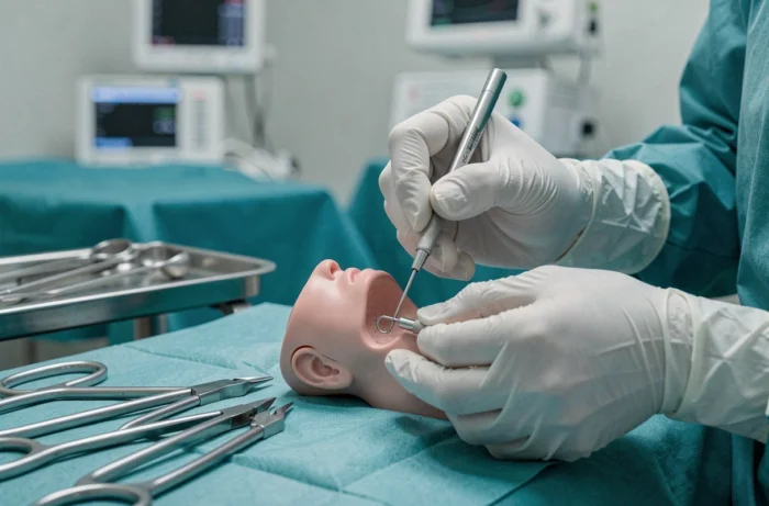
Two conization techniques coexist. The most common uses a diathermy loop (also called LEEP or loop excision). A thin electric wire precisely cuts the tissue. The other method, using a cold scalpel, is reserved for situations where the doctor wants clearer resection margins, for example, in cases of suspected associated glandular lesions.
Why is the analysis of the removed tissue so important? Because it verifies two things: that the lesion has been completely removed (clear margins) and that there was not already a hidden invasive cancer beneath the surface. The treatment of squamous intraepithelial lesions largely relies on this dual diagnostic and therapeutic function of conization.

Active Surveillance of CIN2 in Young Women: A Structured Alternative
Conization is not always the first step. In young women, some lesions classified as CIN2 (a subtype of HSIL) may regress spontaneously. Removing cervical tissue before it is necessary has a concrete drawback: conization can weaken the cervix for future pregnancies, with an increased risk of premature birth.
Several countries have integrated active surveillance as a structured option. National guidelines from Germany, Australia, the UK, and North America provide for this possibility for CIN2 under specific conditions:
- The junction between the two types of cervical cells (transformation zone) must be fully visible during colposcopy.
- There must be no suspicion of invasion or associated glandular lesions.
- The patient agrees to close monitoring, with regular checks through colposcopy and cytology.
- If the lesion persists beyond a defined period, conization treatment is recommended.
Active surveillance does not mean a lack of management. It is a rigorous protocol, with scheduled consultations. If the lesion progresses or persists, the transition to excision is decided without delay.
Thermal Ablation and CO2 Laser: When Destruction is Sufficient
For certain well-defined and fully visible lesions, tissue destruction without sampling may be considered. The CO2 laser vaporizes abnormal cells under colposcopic control. Thermal ablation uses a heated probe to destroy the affected area.
These techniques have an advantage: they preserve more cervical tissue than conization. Their limitation is the lack of a surgical specimen for analysis. Without histological analysis, one cannot exclude a developing cancer. This is why these methods are only proposed after a colposcopy and prior biopsies have already confirmed the absence of invasion.
Thermal ablation is receiving renewed interest in contexts where access to surgery is limited. Recent studies are also exploring the use of local hyperthermia as a complementary approach for precancerous cervical lesions, with still preliminary results.
Laser or Conization: How the Choice is Made
The choice between destruction and excision depends on the size of the lesion, its location, and the diagnostic certainty. When the lesion extends into the cervical canal (endocervix), conization is preferred because the laser cannot safely reach this area. When the lesion is small, superficial, and fully visible, the laser may suffice.
Post-Treatment Follow-Up: What Determines Healing
Treatment does not stop at the intervention. Follow-up after conization or ablation relies on checks combining cytology and HPV testing. The goal is to ensure that the lesion does not recur and that the HPV virus has been eliminated by the immune system.
A negative HPV test after treatment is the best indicator of healing. Conversely, the persistence of the virus after the intervention signals a higher risk of recurrence and justifies enhanced monitoring.
The follow-up schedule varies according to local recommendations, but it generally includes an initial check a few months after the intervention, followed by spaced visits over several years. This extended follow-up is why the vast majority of treated high-grade lesions never progress to invasive cancer.
Regular screening through smears or HPV tests remains the best way to detect these lesions at a stage where they can be easily treated. A high-grade lesion managed in time is cured in the vast majority of cases, whether through conization, structured active surveillance, or laser destruction. The determining factor is not so much the technique chosen but the consistency of gynecological follow-up before and after treatment.